Research teams from Switzerland and Japan have developed a groundbreaking microscopy technique that allows observers to witness the influenza virus entering human cells in real time. This significant advancement provides a clearer understanding of the mechanisms behind viral infections, potentially paving the way for improved antiviral therapies.
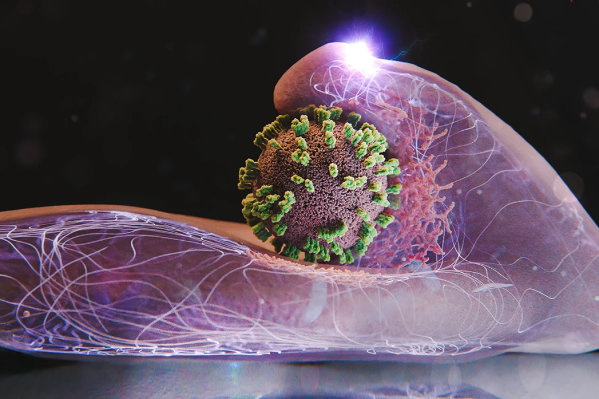
The study reveals how the influenza virus, known for causing seasonal outbreaks, uses two critical proteins—hemagglutinin (HA) and neuraminidase (NA)—to infiltrate host cells. These proteins act as molecular keys, binding to sialic acids on the cell surface, which facilitates the virus’s entry through a process known as endocytosis. Understanding this process is crucial for developing effective treatments against the flu.
Innovative Microscopy Technique Offers New Insights
The newly developed method, termed virus-view dual confocal and atomic force microscopy (ViViD-AFM), combines the capabilities of two advanced imaging techniques. This hybrid system enables researchers to zoom in on living cells with unprecedented detail, allowing them to observe the intricate steps involved in viral entry. The research team, led by Yohei Yamauchi from ETH Zurich, found that the target cells do not passively accept the virus; instead, they actively engage in the encounter.
Using this innovative approach, the researchers documented the dynamic interactions between the flu virus and human cells. They observed how the virus navigates the cell surface, adapting its movements under varying conditions. The team experimented with different viral proteins and binding sites, gaining insights into how the virus modifies its strategy to gain entry.
One particularly striking observation was that the cells respond actively by changing shape and attempting to capture the virus. “The infection of our body cells is like a dance between virus and cell,” stated Yamauchi, highlighting the intricate interplay of forces at work.
Implications for Future Research and Drug Development
The findings, published in the journal PNAS, indicate that influenza viruses require larger protrusions on the cell surface to facilitate entry. These protrusions, composed of the protein actin, play a crucial role in the virus’s ability to penetrate cells. The study also found that once the virus attaches to receptor clusters, it prompts the cell to envelop it in a clathrin coat, effectively pulling the virus further inside.
The ViViD-AFM technique holds significant potential for future research. By enabling scientists to visualize how antiviral drugs affect viral entry in real time, this method can inform the development of more effective treatments. Additionally, the tool’s versatile nature allows for the exploration of other viruses and the interactions between cells and vaccines.
As researchers continue to refine this microscopy technique, it may serve as a valuable “window” into cellular processes. The ability to observe how viruses attach, how cells release extracellular vesicles, and how drug-carrying nanoparticles penetrate cells could lead to major breakthroughs in both biology and medicine.
This advancement in microscopy not only enhances scientific understanding but also brings hope for combating viral infections more effectively, ultimately improving public health outcomes. The collaboration between Swiss and Japanese scientists exemplifies the power of international cooperation in addressing global health challenges.