Researchers at Cornell University have developed a groundbreaking neural implant that is smaller than a grain of salt and capable of wirelessly transmitting brain activity data for over a year. This innovation, detailed in the journal Nature Electronics on November 3, 2023, marks a significant advancement in microelectronics, paving the way for improved neural monitoring and bio-integrated sensing applications.
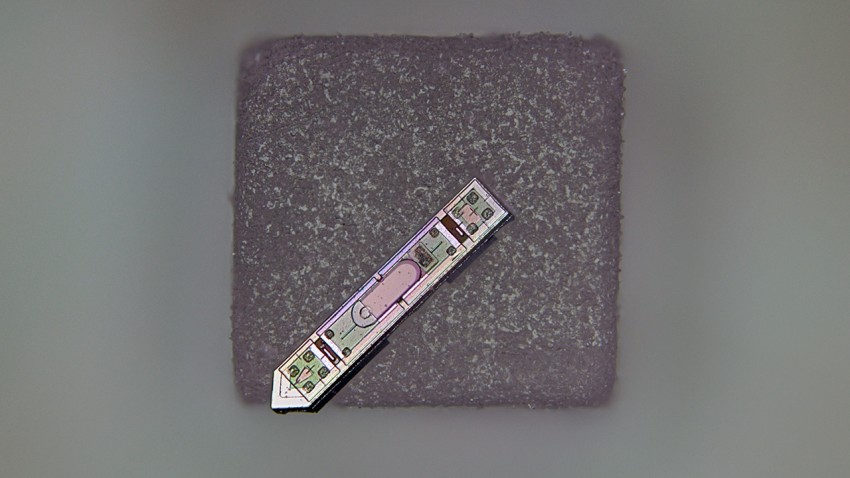
The device, known as a microscale optoelectronic tetherless electrode (MOTE), measures approximately 300 microns in length and 70 microns in width. Co-led by Alyosha Molnar, the Ilda and Charles Lee Professor in the School of Electrical and Computer Engineering, and Sunwoo Lee, an assistant professor at Nanyang Technological University, the MOTE represents the smallest neural implant capable of wireless communication of brain activity.
The MOTE operates using red and infrared laser beams, which can pass through brain tissue without causing harm. It transmits data through tiny pulses of infrared light, encoding the brain’s electrical signals. A semiconductor diode made from aluminum gallium arsenide captures light energy to power the device and emits light to relay information. The MOTE is equipped with a low-noise amplifier and optical encoder developed from the same semiconductor technology found in everyday microchips.
Alyosha Molnar stated, “As far as we know, this is the smallest neural implant that will measure electrical activity in the brain and then report it out wirelessly.” The implant utilizes pulse position modulation, a technique also employed in optical communications for satellites, allowing it to function efficiently with minimal power consumption.
The research team initially tested the MOTE in cell cultures before implanting it into the barrel cortex of mice, the area of the brain responsible for processing sensory information from whiskers. Over the course of a year, the implant successfully recorded both spikes of electrical activity from neurons and broader patterns of synaptic activity, all while keeping the mice healthy and active.
Molnar noted that traditional electrodes and optical fibers can irritate brain tissue, potentially triggering an immune response. The design of the MOTE aims to minimize this disruption while capturing brain activity more rapidly than imaging systems, and without the need for genetic modifications to neurons.
Looking to the future, the MOTE’s material composition could allow for electrical recordings from the brain during MRI scans, a feat currently unattainable with existing implants. The technology also has the potential to be adapted for use in other tissues, such as the spinal cord, and could even be integrated with innovations like opto-electronics embedded in artificial skull plates.
The concept for the MOTE was first conceived by Molnar in 2001, but significant progress occurred after he began discussions with members of Cornell Neurotech, a collaborative initiative between the College of Arts and Sciences and Cornell Engineering, approximately ten years ago. The study’s co-authors include Chris Xu, director of the School of Applied and Engineering Physics; Paul McEuen, the John A. Newman Professor Emeritus in the Department of Physics; Jesse Goldberg, the Dr. David Merksamer and Dorothy Joslovitz Merksamer Professor in Biological Sciences; and Jan Lammerding, professor in the Meinig School of Biomedical Engineering.
This research was partially funded by the National Institutes of Health, with fabrication work conducted at the Cornell NanoScale Facility, which receives support from the National Science Foundation. As highlighted by Syl Kacapyr, associate director of marketing and communications for Cornell Engineering, this research opens new avenues for how we understand and monitor brain activity in living organisms.