Colorectal liver metastases (CRLM) represent a significant challenge in cancer treatment, often evading conventional therapies. Researchers from the biomedical engineering department at Texas A&M University have made strides in addressing this issue by developing advanced models and imaging techniques aimed at detecting and understanding this aggressive form of cancer.
CRLM occurs when cancer spreads from the colon or rectum to the liver, where it can hide in the complex architecture of the organ. Standard treatments, such as chemotherapy and surgical resection, frequently fail, with as many as 75% of patients experiencing recurrence within two years, leading to poor survival rates. This high rate of recurrence is partly due to cancer cells taking refuge in the liver’s intricate ridges and folds, making them difficult to detect and treat effectively.
A team led by Dr. Shreya Raghavan, along with colleagues Dr. Alex Walsh and Dr. Daniel Alge, has dedicated their research to illuminating these hidden cancer cells. Their findings, published in the journal Advanced HealthCare Materials and Biomaterial Sciences, highlight the challenges of visualizing dormant cancer cells. “The thing with dormant, hidden cancer is that it is really hard to detect,” Raghavan stated. Collaborating with experts from MD Anderson Cancer Center, the researchers noted that conventional imaging techniques often fail to identify these tumors.
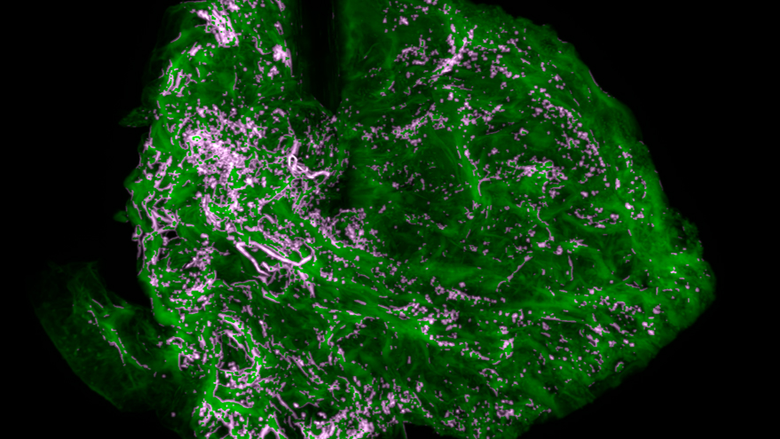
In a breakthrough, Dr. Sabrina VandenHeuvel, a co-author of the study and former graduate researcher in Raghavan’s lab, developed a method to grow colon cancer cells on a liver scaffolding material. This innovation allowed the team to replicate the dormancy effect observed in patients. “This is a pretty big deal. We can engineer a model of dormant colon cancer liver metastasis,” Raghavan explained. This model enables researchers to explore unique characteristics of the disease that are often undetectable in clinical settings.
Despite their initial success, the researchers faced limitations with traditional microscopy methods when trying to image the metastases. Dr. Oscar Benavides, a co-author and instructional assistant professor, introduced a new imaging technique capable of providing higher spatial resolution. “The microscope that I use illuminates only a very thin plane, like five micrometers thick, which gives us really high spatial resolution,” Benavides noted. This method allows for three-dimensional reconstructions, enhancing the ability to visualize structures beyond the reach of standard techniques.
This research project received funding through the National Institutes of Health’s R37 MERIT program, underscoring its significance in advancing cancer research. The team has also created an open-source imaging and machine learning platform, making it available for other researchers to adapt for their own studies. “We see this as a proof of concept to show we can generate these metastatic cancer models,” Benavides said. By sharing their resources, the researchers encourage collaboration within the scientific community to further investigate CRLM.
Looking ahead, Raghavan’s lab plans to collaborate with Alge’s lab to enhance liver scaffolding systems and gain insights into factors that contribute to cancer dormancy. Concurrently, Walsh’s lab is working on refining microscopy techniques to identify potential treatments for the metastases. “We all play distinct roles in this bigger interdisciplinary project,” Benavides explained. “By understanding how cells evade detection and recurrence, doctors could one day match the right drug to the patient’s unique metastatic disease, improving outcomes for the thousands facing colorectal liver metastases each year.”
The collaborative efforts at Texas A&M University reflect a commitment to advancing cancer treatment and improving patient outcomes, addressing a critical need in the field of oncology.